TECHNICAL BACKGROUND
THE HIGHEST-QUALITY INFRASTRUCTURE IN THE MILITARY UNIVERSITY OF TECHNOLOGY BIOMEDICAL ENGINEERING CENTRE.
The Military University of Technology Biomedical Engineering Centre conducts its research on graphene, using the highest-quality infrastructure. At its disposal, the centre has modern equipment and test apparatus in five specific laboratories, which conduct research and implementation.
Molecular Engineering and Graphene Laboratory is one of the laboratories currently conducting the research concerning, among others, the development of novel technologies of merging graphene with other materials, the processes of producing molecular sieves and active adhesive surfaces for biological particles. Moreover, it works on a simplified laboratory device for one-stage enrichment of circulating tumour cells (CTC) in the blood samples.
In order to perform the above-mentioned tasks, the Laboratory operates laboratory devices for the production of molecular sieves, along with technological instrumentation. Those devices are required for performing direct laser photolithography. They include devices for ion etching, plasma cleaning, forming active surfaces and geometric control of the produced molecular sieves.
Research infrastructure of the Molecular Engineering and Graphene Laboratory
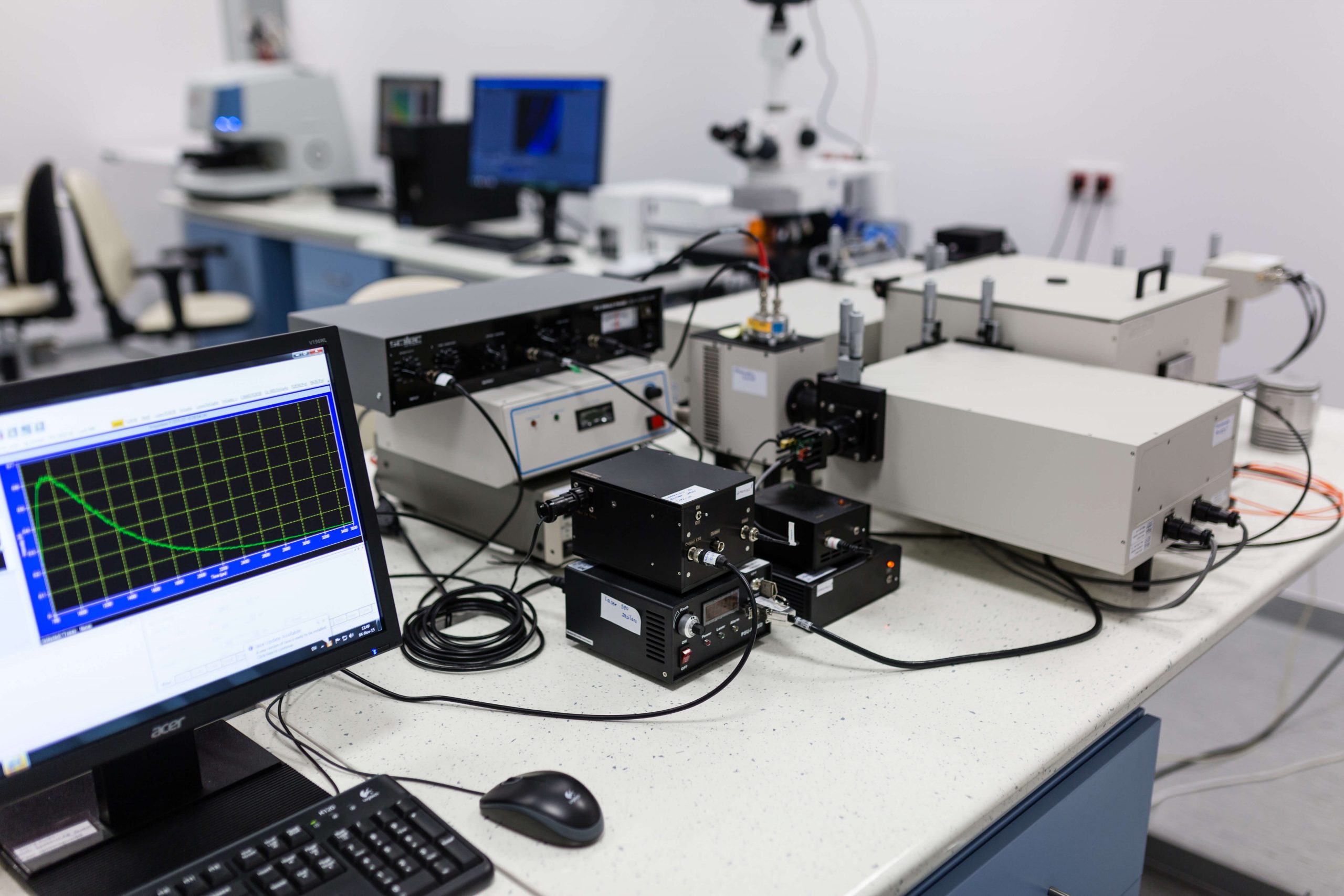
Spectrofluorometer
Type: Horiba QM 400
Quanta Master QM400 spectrofluorometric system is designed for the specification of luminescent materials in the form of powders, solutions, thin layers, crystals and nanomaterials. The spectrum of excitation and detection lies between 200 and 2200nm. Excitation is provided by: NA: YAG Opolette 355LD (225-2200nm) impulse laser, ellipsoidal reflector with Xe lamp (200-900nm) and 980nm laser for measuring up conversion. The system allows measuring excitation, emission, synchronic, as well as non-synchronic spectrums, fluorescence kinetics and anisotropy of luminescence polarisation. The measurements are taken with the use of absorption and transmission attachment, as well as of integrating sphere. The system is conjugated with a fluorescence microscope (Zeiss Axio Imager.M2) and a super-sensitive video recorder for measuring bioluminescence.
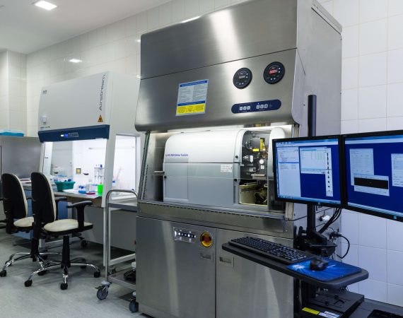
Multilaser cell sorter
Type: BD FACSAria Fusion fusion flow cytometer
The sorter is integrated with class II type A2 biosafety cabinet. It also has AMS system (Aerosol Management System), which protects users from being exposed to aerosols created when using the device. BD FACSAria Fusion enables a quick quantitative and qualitative assessment of physical and biological properties of single cells and their morphotic elements, such as nuclei, nucleic acids or mitochondria. The device can have various applications, including the assessment of the expression ofsurface and intracellular antigens, determination of the cell cycle phase, analysis of cytoplasmic membrane properties, isolation of cells very rarely encountered in a population, as well as studying apoptosis, necrosis and activity of enzymes. The device may also be used for the aseptic sorting of single cells, for example, for the purposes of cloning.
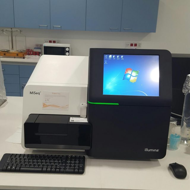
Genome sequencer
Type: Illumina Inc. MiSeq with additional accessories: Promega Quantum fluorometer, Hybex heating bloc, BluePippin device for the electrophoresis of nucleic acids and proteins, computer system with NAS disc memory for analysing and storing the results of sequencing with the use of a dedicated Geneious RS software.
The sequencer is designed for the analysis of the genetic material in mammal or microbial cells by targeted sequencing of the genome fragments with the presence of mutations related to specific diseases, metagenomics, for example, identification of numerous species of microbes/pathogens in a test tube, sequencing of small genomes, miRNA sequencing, targeted gene expression, amplicon sequencing, compiling a database for NGS (next-generation sequencing), for example, for the purposes of whole genome DNA methylation analysis, detecting mutations related to specific types of tumours in a gene/sequence pool, detecting remaining tumour cells (minimal residual disease) which might cause tumour recurrence, assessment of post-transplant chimerism (graft acceptance assessment).
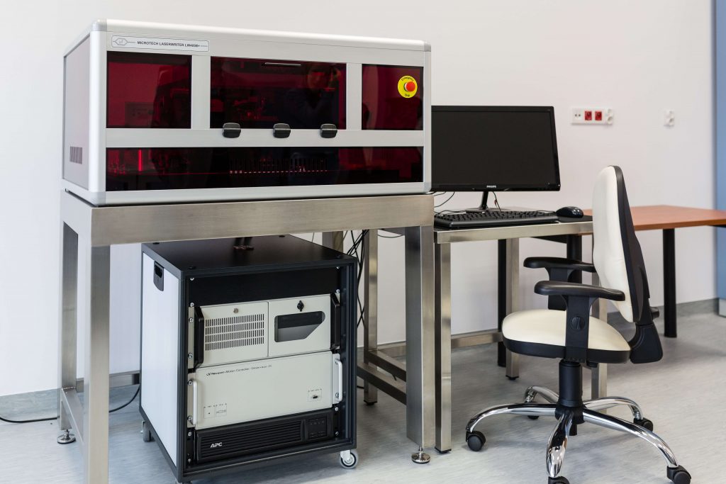
Direct laser photolithography device
Type: MicroTechs. r. l. LaserWriter 405B+
LaserWriter is a direct laser photolithography device, is a necessary apparatus for forming high-resolution geometric patterns with 1.0 micrometre resolution on metal and semiconductor surfaces used for ion etching (DRIE). It constitutes standard equipment for producing molecular sieves for CTC enrichment. In comparison with standard photolithography, this is a more flexible and much cheaper method. Moreover, it enables a rapid generation of specific high-resolution patterns required for the purposes of the ion etching of molecular sieves.
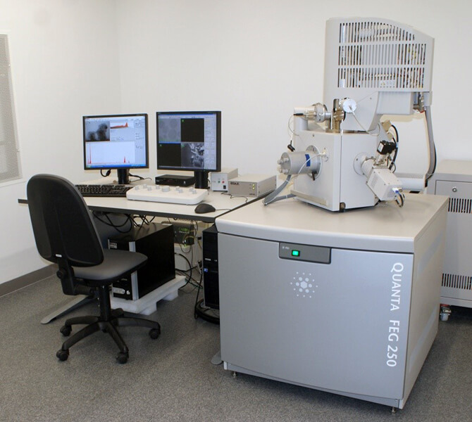
High-resolution scanning electron microscope with EDS spectrometer and a complete set of equipment for analysing biological samples
Type: FEI Quanta 250 FEG
Quanta 250 FEG high-resolution scanning electron microscope (SEM – Schottky emitter) with EDS spectrometer and a complete set of equipment for imaging biological samples is dedicated for quantitative and qualitative assessment of CTC filtration process, conducted using molecular sieves and a prototype device for blood screening test. Quanta 250 FEG allows biological samples to be compared, using a much lower accelerating voltage, in comparison with tungsten filament or LaB6 filament, still used by a small number of manufacturers. Quanta microscope is an environmental microscope, and therefore is most suitable for imaging samples which are incompatible with traditional electron microscopy (biological, porous, hydrated samples etc.). Peltier cooling stages allow for the regulation of environment humidity of an imaged substance via regulation of temperature and pressure in the chamber.Moreover, the device has an extensive low vacuum system, allowing the pressure in the chamber to drop to 4000 Pa, as well as a number of safety systems protecting the microscope from any kind of pollution. The microscope is equipped with EDS spectrometer, along with software and a computer suitable for data processing. The set also contains SE detectors and unique solutions for BSE signal detection.
HISTORY
- Discovery of graphene properties – the first isolation of graphene layer (2004),
- The Nobel Prize awarded for the discovery (2010),
- Commencement of the research on graphene—based antibacterial paper as dressing material (2010),
- Founding of the Biomedical Engineering Centre Cluster —CIBio WAT (2012),
- Granting funds for POIG (Innovative Economy Operational Programme) to the Warsaw Medical University, leading CIBio WAT to become the only cluster in Poland to be coordinated by a higher education institution (2013),
- Positive results of cytotoxicity tests of graphene oxide. Research was conducted in Warsaw Medical University Department of Transplantology and Central Tissue Bank. The research was concluded by positive results and further research was recommended (2014),
- Confirmation of antibacterial properties of graphene oxide by CIBio WAT (2015),
- Patent application for a cosmetic composition containing graphene as a bactericide P.424202 (2018),
- Confirmation of the assumption that the fibrous structure of the gel can be strengthened by embedding nanostructures (2019),
- Successful trials of embedding graphene and graphene oxide flakes into the structure of the gel, which means that we have concluded laboratory tests with satisfying results (2019),
- Optimisation of the graphene/gel composite – laboratory-scale technology (2019),
- Application tests and technology scaling (2020),
- Testing functionality of graphene oxide flakes – the optimisation of the mechanism of embedding graphene into the structure of the gel and surface regeneration of tissue structures using bioactivity (2019),
- Optimisation of the graphene / gel composite – laboratory-scale technology (2019),
- Application tests and technology scaling (2020),